Facts & figures
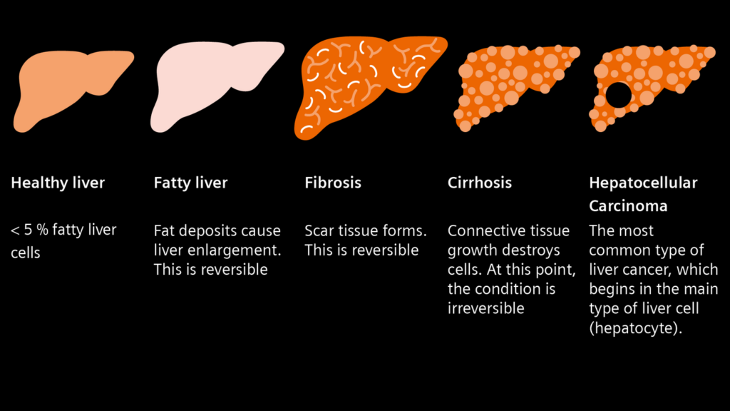
A healthy liver is able to perform its normal functions effectively, for example aiding digestion and breaking down harmful drugs and poisons. Continuous inflammation of the liver can lead to fibrosis – a formation of scar tissue within the liver. And extensive scarring can block the flow of blood through the liver and cause liver function to deteriorate over time. This is called cirrhosis and may eventually lead to hepatocellular carcinoma (HCC).
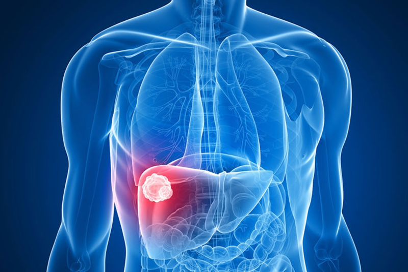
Several types of cancer can form in the liver. The most common type of liver cancer is hepatocellular carcinoma (HCC), which begins in the main type of liver cell (hepatocyte). Other types of liver cancer, such as intrahepatic cholangiocarcinoma and hepatoblastoma, are much less common.
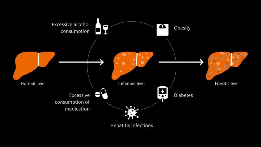
There are several factors that can lead to liver disease. Excessive alcohol consumption, obesity, diabetes, hepatitis infections, and excessive consumption of medication can all contribute to an inflamed liver and tissue damage.
A silent killer
Over 90 percent of cases of hepatocellular carcinoma (HCC) occur in patients with chronic liver disease. It is often described as a “silent killer” as patients can be at risk of death even without exhibiting symptoms. The signs and symptoms of liver cancer are most often the result of liver damage and may include yellowing of the skin, right-sided abdominal or shoulder blade pain, or a lump in the right upper abdomen. However, many of the warning signs are non-specific, such as weight loss and fatigue.
The liver does not have pain receptors. Patients cannot feel inflammation. This means the fibrosis of the liver usually remains undetected and early diagnosis is a challenge. Strictly speaking, fibrosis is not a disease in its own right as it only occurs as a symptom of other diseases. Its gradual progression is critical because medical research has previously assumed that scarring of the liver is irreversible once it has become advanced.
Diagnosis
Liver disease can be diagnosed in various ways. As the scar tissue accumulates, the liver loses some of its elasticity and becomes stiffer. An elastography or ELF test can help diagnose and monitor liver disease or damage.
The ELF test
A blood sample is taken. Three important serum markers can be detected with an automated analyzer and the risk of disease progression can be derived from these.

Elastography
A probe emits a mechanical pulse toward the liver. An integrated ultrasound transducer measures the velocity of the pulse wave between two points. The less elastic the liver tissue, the faster the pulse propagates through the liver.

Biopsy
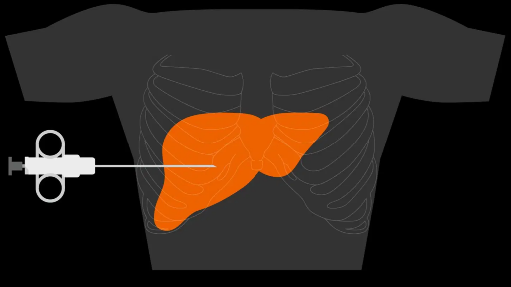
A tissue sample is taken from the liver with a cannula. The sample is then examined for scar tissue under a microscope.
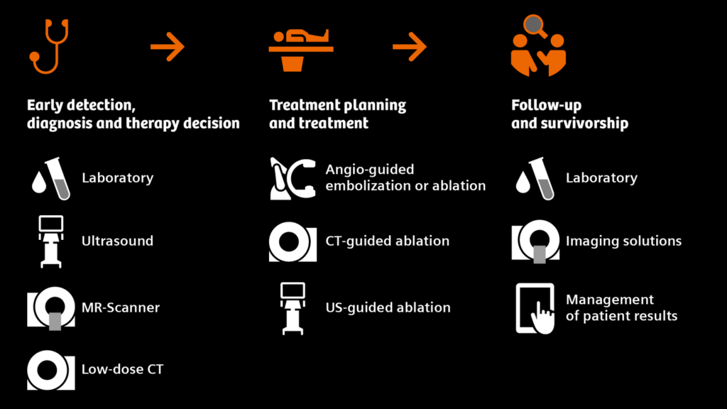
By contrast, diagnosing liver cancer requires a physical examination and special medical tests. Diagnosis of hepatocellular carcinoma (HCC) is done using imaging tools like ultrasound, computed tomography (CT), or magnetic resonance imaging (MRI) and in many cases doesn’t even require biopsy.
Siemens Healthineers provides imaging technology and the smart imaging value chain that enable reliable therapy decisions. Sometimes a liver biopsy is performed to confirm the diagnosis of liver cancer. Genetic testing of the cancer can help determine the best type of treatment for the patient.


Write a comment
Your email address will not be published. All fields are required